Dr. Pushkar Lele, assistant professor in the Artie McFerrin Department of Chemical Engineering at Texas A&M University, has received a grant from the Department of Defense (DOD) – Army Medical Research and Materiel Command. The project will determine how bacteria form complex, macromolecular apparatuses, and how environmental factors trigger their assembly.
Bacteria, like most living systems, rely on molecular machines to do just about everything from the synthesis of enzymes, DNA replication, and intracellular signaling, to surface-colonization, cell division
According to Lele, the central question driving the research is how the kinetics of self-assembly influence bacterial adaptation.
“How quickly can a particular machine be built to respond to an environmental stress,” asked Lele? “That would determine whether it is of any use – it is not adequate to be merely effective in function, the rate of assembly is likely to be important, especially under duress. We are proposing to study this by introducing an environmental stressor – it could be chemical, it could be thermal, it could be mechanical – and asking, at what rate will the assembly progress and how will that impact the chances of survival.”
Ultimately, Lele hopes to learn the principles by which environmental cues can guide cellular self-assembly, with potential future applications in biomimetics and nanotechnology.
To study the effects of environmental stressors on intracellular self-assembly, Lele and his research team will employ a novel combination of three different techniques. The first is total internal reflection fluorescence microscopy (TIRF). This technique allows researchers to follow the dynamics of protein molecules within the cell. In order to visualize these proteins, Lele's group generates and employs fusions of the proteins of interest with a protein that fluoresces when illuminated by a powerful green laser.
The second technique is called optical tweezers. It involves the use of a highly focused laser beam to indefinitely trap and hold microscopic objects at desired locations. Optical tweezers were invented by Arthur Ashkin in 1986, who recently won a Nobel Prize in Physics for his work on the technique.
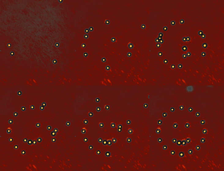
“Focusing the laser results in intense nonuniform electric fields,” Lele said. “Under the right conditions, it helps trap di-electric particles, such as bacterial cells. A cell will remain trapped in one position and will go wherever the laser beam is moved within the sample plane. By rapidly scanning the laser between multiple positions, multiple bacterial cells can be trapped simultaneously.”
Lele and his team have now combined optical tweezers with TIRF, enabling them to translate bacteria toward or away from a stressor, as desired, while observing the intracellular self-assembly process.

The third technique is called Förster resonance energy transfer (FRET). In this technique, two types of fluorescent fusions are employed that form a ‘FRET pair.’ When the two fluorophores are within a short distance under illumination, a nonradiative transfer of energy occurs resulting in emissions at certain wavelengths of light. These can be detected with a photomultiplier or a sensitive camera. As a result, FRET has become an important tool for in vivomeasurements of protein-protein interactions. Lele hopes to use FRET in combination with the above two methods to develop a comprehensive understanding of the importance of timing in the self-assembly and functioning of protein complexes.
“It is ambitious, but it promises powerful insights into the cellular self-assembly process,” Lele said, adding that the survival of bacteria depends on the exquisite timing of a variety of important events. “We are trying to understand how some of the events are precisely timed to give rise to protein machines.”