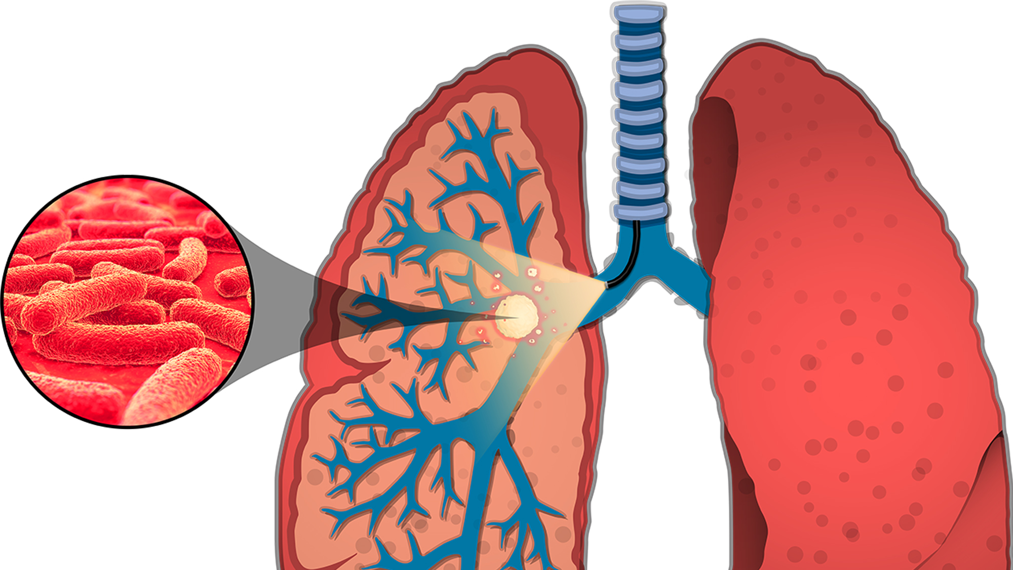
Tuberculosis is a highly contagious and deadly disease that has recently surpassed the human immunodeficiency virus (HIV) in number of worldwide deaths. Current methods of diagnosing and treating tuberculosis take time and are not always successful, leading to wider proliferation of the disease. Researchers at Texas A&M are working to create more efficient and effective diagnostic tools.
Dr. Kristen Maitland, associate professor in the Department of Biomedical Engineering, is working with Dr. Jeffrey Cirillo, director of the Center for Airborne Pathogen Research and Tuberculosis Imaging and professor in the Department of Microbial Pathogenesis and Immunology, on the project, funded by the National Institute of Allergy and Infectious Diseases and the National Science Foundation.
Using optical imaging technology, Cirillo and his team developed a method of observing bacteria in a living animal’s lungs without harming them. The scan lights up the bacteria in the lung through fluorescence-based technology. When Maitland joined the team, she brought an innovation that would allow for an even more detailed result, a micro-endoscope that could be inserted into the airway and detect smaller populations of bacteria in the lungs.
“It is a much more sensitive approach,” Maitland said. “Using the micro-endoscope to light up the lungs from the inside has resulted in 100-1000 times improvement in the detection of pulmonary infection.”
Using this technology, researchers can study airways in both animals and humans to detect and diagnosis tuberculosis much sooner than current methods, which can lead to better treatments.
Along with the probe, Maitland and her team have developed computer models of the lungs that enable them to test the fluorescence-based equipment on a variety of simulated species, even young children. They have also used 3D printing to develop a phantom tissue that looks like an airway to light and can be used to improve the effectiveness of the light-based probe.