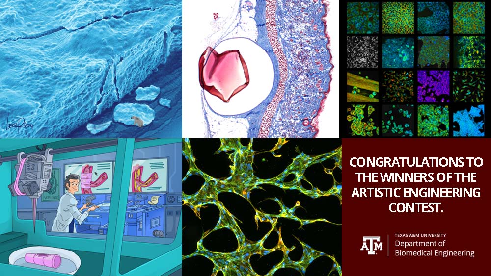
The Department of Biomedical Engineering at Texas A&M University hosted its first annual Artistic Engineering art contest during the fall 2021 semester. Biomedical engineering-affiliated students, faculty and staff were invited to submit images in any medium and could include themes such as research, people in STEM, computer-generated imagery, photo composites or others.
Voting took place and the top five submissions were selected. These will be featured on the fifth floor of the Emerging Technologies Building.
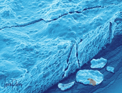
First place: Laura Rivera Tarazona, doctoral student in Dr. Taylor Ware's lab, for
“Living Materials Save the Environment.” The glacier presented in this piece is an scanning electron microscope’s image of engineered living materials (ELM). The ELM material in this image consists of Escherichia coli contained within a hydrogel matrix. In the Ware Lab, they are synthesizing these ELMs to replace traditional plastics and help the global sustainability of natural environments.
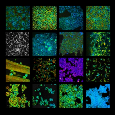
Third place: Members of the Quantitative Optical Imaging Lab led by Dr. Alex Walsh for
“Fluorescence Collage,” representative label-free fluorescence images of cell metabolism from the Quantitative Optical Imaging Lab. Fluorescence lifetime imaging is used to quantify the amount of time a fluorophore is in an excited state, which can enhance our understanding of certain microenvironmental changes. Contrast comes from molecules within cells, nicotinamide adenine dinucleotide (NADH) or flavin adenine dinucleotide (FAD), which are energy carriers used in metabolic reactions within the cell. Collage contains images of cancer cells, liver tissues, macrophages and organoids.